A nuclear medicine scan is an imaging test to create pictures of your organs and bones. It uses a special camera and a small amount of radioactive material called a radiotracer.
Why is a nuclear medicine scan done?
A nuclear medicine scan can create pictures of your heart, lungs, liver, and gallbladder. Nuclear medicine scans help healthcare providers diagnose health conditions such as cancer, injuries, and infections. They can also show how well the organs like your heart and lungs are working. Some common types of nuclear medicine scans include:
-
PET scan (positron emission tomography scan)
-
Specific body part scans (such as bone scans, biliary scans, thyroid scans, and cardiac scans)
-
SPECT (single photon emission computed tomography) scan
-
PET/CT scans (PET/computerized tomography scans)
-
MUGA (multi-gated acquisition) scan
-
Gallium scan
Risks of a nuclear medicine scan
Nuclear medicine scans are largely safe. The dose of radiation in the tracer is quite small. It is generally not a cause for serious concern. The risk of toxic or allergic reactions with tracers is also low. Some people may get pain or swelling at the site of injection. In rare cases, allergic reactions may occur in some people.
Before the scan
The way you get ready for a nuclear medicine scan depends on the type of scan you will have. Follow your healthcare provider's instructions.
-
Tell your provider about all the medicines you take. Ask if it’s OK to take them before your test. This includes prescription and over-the-counter medicines. It also includes vitamins, herbs, and other supplements.
-
Follow all instructions you are given for not eating or drinking before the test. You may be told not to have caffeine or tobacco for a certain amount of time before the test.
-
Your scan may take a few hours. Bring something you can do if you need to wait.
-
You will be given a radioactive tracer. It may be injected, swallowed, or inhaled. If injected, you will get an IV (intravenous) line. Your scan may then be done right away. Or you may need to wait a few hours or even days to allow the tracer to concentrate in the part of the body being studied. You may be scanned many times during 1 day. This depends on the type of nuclear scan you have.
Tell the technologist
Tell them if you:
-
Are pregnant or think you may be
-
Are breastfeeding (You may need to pump breastmilk before the procedure. You may need to discard breastmilk until the tracer is gone from your body.)
-
Had a nuclear medicine scan before
-
Had a recent barium study or an X-ray using contrast
-
Have any breaks (fractures) or artificial joints
-
Have any allergies
During the scan
-
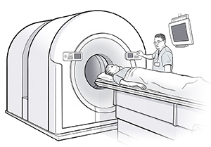
You will lie on a narrow imaging table.
-
A large camera is placed close to your body.
-
Stay as still as you can while the camera takes the pictures. This will ensure the best images.
-
The table or camera may be adjusted to take more pictures.
After the scan
-
Most of the radioactive tracer will leave your body through pee and poop in a few days. Drink plenty of water to help clear the tracer from your body. After using the toilet, put the lid down and flush the toilet right after you use it.
-
Your healthcare provider will discuss the test results with you during a follow-up visit or over the phone.
-
Some of the tracer in your body will naturally lose its radioactivity with time. Talk with your healthcare provider about any steps you need to take with sex, or being close to pregnant people or children after the test.


